WELCOME to the homepage of Prof. Dr. Silvio O. Rizzoli
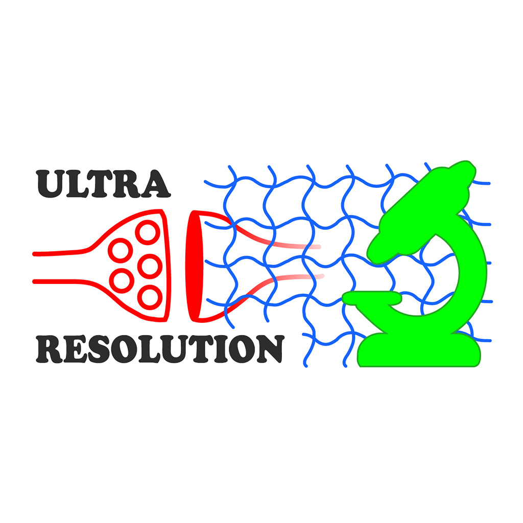
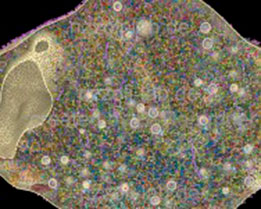
The diffraction barrier has been particularly problematic for imaging synaptic vesicles, which are among the smallest known organelles (30-50 nm in diameter). They are located in small areas in the synapses (about 1 micron in diameter).
Our group takes advantage of increase imaging resolution provided by different types of microscopy to investigate synaptic vesicle function, with an emphasis on synaptic vesicle recycling. Since STED microscopy also allows imaging of protein domains, the group aims at studying the patterning of protein domains in the synapse, in order to understand its molecular architecture.